Pulmonary Medicine
Medically reviewed by Varun Halani, MD · August 13, 2026
Interstitial Lung Disease (ILD)
An overview of interstitial lung disease as an umbrella term for over 200 conditions that scar or inflame the lung interstitium, how it differs from idiopathic pulmonary fibrosis, and how it is diagnosed and managed.
In short
Interstitial lung disease (ILD) is an umbrella term for more than 200 conditions that scar or inflame the interstitium, the supportive tissue around the lungs' air sacs. It is not another name for idiopathic pulmonary fibrosis (IPF), which is one specific ILD subtype among many. Diagnosis combines history, exposure review, pulmonary function testing, and HRCT imaging, often with a multidisciplinary discussion, and treatment depends on the specific subtype identified, ranging from antifibrotic therapy to anti-inflammatory treatment to removing an identified exposure.
Interstitial Lung Disease (ILD) at a Glance
What it is
An umbrella term for more than 200 conditions that scar or inflame the interstitium, the supportive tissue around the lungs' air sacs.
Not the same as IPF
Idiopathic pulmonary fibrosis is one specific ILD subtype, not another name for the whole category.
Common early symptoms
Exertional shortness of breath and a persistent dry cough that develop gradually.
Key test pattern
A restrictive pattern on pulmonary function testing, distinct from the obstructive pattern in asthma or COPD.
Diagnostic approach
History, exposure review, pulmonary function testing, HRCT imaging, selected blood tests, and often a multidisciplinary discussion.
Treatment approach
Depends on subtype, ranging from antifibrotic therapy to anti-inflammatory treatment to removing an identified exposure.
Key Takeaways
- Interstitial lung disease (ILD) is an umbrella term for more than 200 distinct conditions that scar or inflame the interstitium, the supportive tissue around the lungs' air sacs.
- ILD is not the same thing as idiopathic pulmonary fibrosis (IPF). IPF is one specific, well-defined subtype of ILD, not a synonym for the whole category.
- ILD causes a restrictive breathing pattern on pulmonary function testing, which is a different mechanical problem than the obstructive pattern seen in asthma or COPD.
- Getting an accurate ILD diagnosis often requires a multidisciplinary discussion among pulmonology, radiology, and sometimes rheumatology and pathology, because different subtypes can look alike on any single test.
- Treatment is subtype-specific: antifibrotic medications target progressive fibrotic patterns like IPF, while anti-inflammatory or immunomodulatory approaches are more relevant for many connective-tissue-disease-associated forms.
- Progressive exertional shortness of breath and a persistent dry cough are the most common presenting symptoms, though the underlying cause and expected course vary widely by subtype.
Symptoms
Respiratory Symptoms
- Shortness of breath that starts with exertion, such as climbing stairs or walking briskly, before it is noticeable at rest
- A persistent dry cough that lingers for weeks or months without an obvious explanation such as a recent infection
Other Possible Symptoms
- Fatigue
- Joint pain or skin changes, when an underlying autoimmune condition is present
Could This Be ILD?
Why am I short of breath doing things that used to be easy?
In ILD, shortness of breath typically develops gradually and shows up first during exertion, such as climbing stairs or walking briskly, before it is noticeable at rest. Because this pattern is common to many other conditions, it is sometimes attributed to aging or deconditioning before ILD is considered.
Why will my dry cough not go away?
A persistent dry cough that lingers for weeks or months without an obvious explanation, such as a recent infection, is one of the hallmark early symptoms of ILD.
I have an autoimmune condition. Could my breathing symptoms be related?
ILD can develop as a complication of an autoimmune or rheumatologic condition, such as rheumatoid arthritis, systemic sclerosis (scleroderma), or inflammatory myopathies. In these cases, the lung disease is closely tied to the underlying autoimmune process and is often managed alongside it.
My chest imaging showed something unexpected. What does that mean?
Abnormal findings on chest imaging obtained for another reason are a recognized reason to pursue further evaluation for ILD, since such findings can be present before symptoms are pronounced enough to prompt a workup on their own.
Major categories of ILD
Idiopathic interstitial pneumonias (including IPF)
A group of ILD subtypes without a single identifiable external cause. Idiopathic pulmonary fibrosis, a progressive scarring disease, belongs here as one specific pattern among several.
Connective-tissue-disease-associated ILD
ILD that develops in the context of an autoimmune or rheumatologic condition, such as rheumatoid arthritis, scleroderma, or inflammatory myopathies.
Exposure-related disease
ILD linked to occupational or environmental exposures, including certain organic dusts, molds, or bird proteins (hypersensitivity pneumonitis) and mineral dusts (pneumoconiosis).
Medication-related ILD
A recognized but less common cause in which certain medications trigger lung inflammation or scarring, identified by reviewing the full medication history.
Other and unclassifiable forms
Less common categories, including granulomatous ILD and cases where the specific pattern cannot be confidently classified despite thorough evaluation.
Risk Factors
- A diagnosed autoimmune or connective tissue diseaseConditions such as rheumatoid arthritis, systemic sclerosis (scleroderma), or inflammatory myopathies are linked to connective-tissue-disease-associated ILD.
- Occupational or environmental exposuresOrganic dusts, molds, and bird proteins can cause hypersensitivity pneumonitis; certain mineral dusts, often in occupational settings, can cause pneumoconiosis.
- Certain medicationsA number of medications, used for various medical conditions, are recognized but relatively uncommon causes of ILD.
- A family history of lung diseaseReviewed as a standard part of the history when ILD is being considered.
How ILD Affects Daily Life
Exercise Tolerance and Daily Energy
Some people with a mild or stable form of ILD continue most of their usual activities with only minor adjustments, while others notice a more significant effect on exercise tolerance and daily energy.
Oxygen Levels During Exertion
When exertional oxygen levels are affected, daily impact tends to be more noticeable, and supplemental oxygen during activity may be appropriate.
Respiratory Infection Risk
Respiratory infections can have an outsized effect on lung tissue already affected by ILD, which is why staying current on routine vaccinations and promptly addressing new or worsening symptoms is generally sensible.
Pulmonary Rehabilitation
Supervised exercise training combined with breathing-technique and energy-conservation education can improve exercise capacity and quality of life even though it does not directly change the underlying disease process.
When Should I Talk to a Pulmonary Specialist?
- Gradually worsening shortness of breath with activity that has been present for more than a few weeks
- A dry cough that persists without a clear explanation such as infection or reflux
- A known autoimmune or connective tissue condition combined with new breathing symptoms
- A history of occupational, environmental, or medication exposures linked to lung scarring
- Abnormal findings on chest imaging obtained for another reason
What “interstitial lung disease” actually means
What ILD Affects
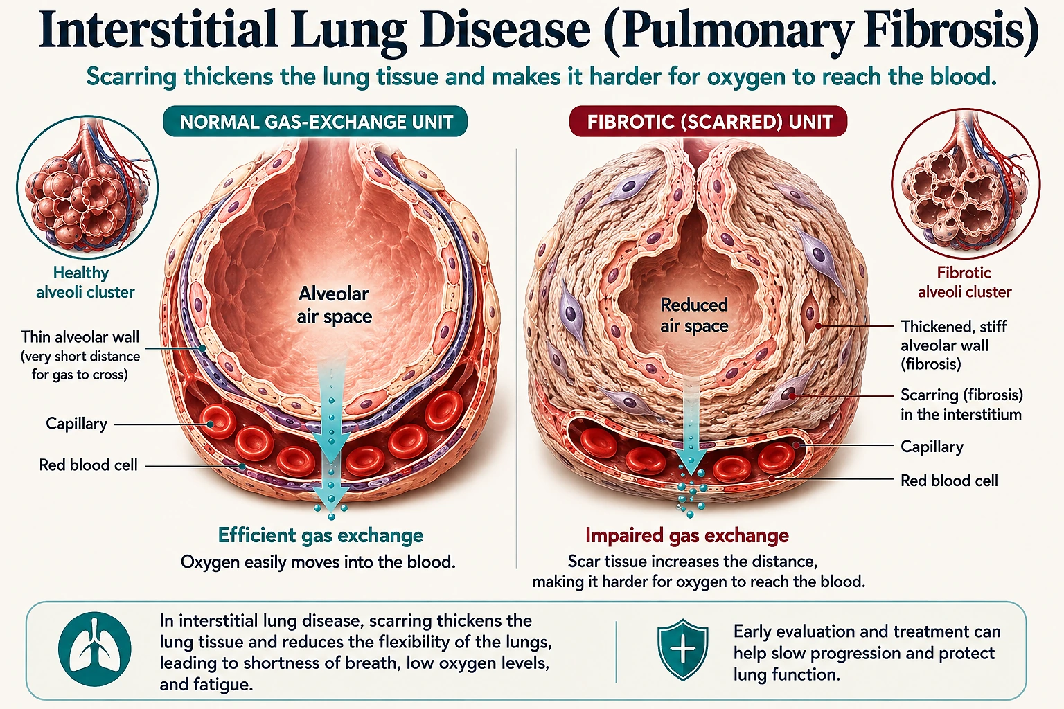
Interstitial lung disease, or ILD, is not one illness. It is an umbrella term that covers more than 200 distinct conditions that share a common feature: they cause inflammation, scarring, or both in the interstitium, the thin layer of supportive tissue and small blood vessels that surrounds the lungs’ millions of tiny air sacs, or alveoli.
This is different from the airways themselves, the tubes that carry air in and out of the lungs and the primary site of disease in conditions like asthma and COPD. In ILD, the problem sits in the tissue and space around the air sacs, which stiffens the lung and can interfere with how efficiently oxygen moves from inhaled air into the bloodstream.
Why the Category Distinction Matters
Because ILD is a category rather than a single diagnosis, two people who are both told they have “interstitial lung disease” can have meaningfully different conditions, different causes, different expected courses, and different treatments.
Getting the specific subtype right is one of the central goals of ILD evaluation. It shapes everything that follows, from testing to treatment to monitoring.
ILD is not the same thing as IPF
This is the single most important distinction to understand about ILD, and it is also the most common point of confusion. Idiopathic pulmonary fibrosis, or IPF, is a specific, well-defined subtype of ILD: a progressive scarring disease of unknown cause with a particular pattern on imaging and, when needed, on tissue sampling.
IPF is one of the better-known and more extensively studied forms of ILD, which is part of why it sometimes gets used loosely as a stand-in for the entire category. But IPF is not a synonym for ILD as a whole, and treating it that way can lead to real misunderstandings about prognosis and treatment.
Many other forms of ILD are quite different from IPF, as the comparison below outlines.
IPF vs. Other Forms of ILD
| Dimension | Idiopathic Pulmonary Fibrosis (IPF) | Many Other Forms of ILD |
|---|---|---|
| Underlying Process | Progressive scarring, or fibrosis | Often active inflammation that can improve substantially with treatment rather than irreversible scarring |
| Cause | Idiopathic, meaning of unknown cause | Often an identifiable trigger, such as an autoimmune condition or an inhaled exposure |
| Typical Course | Tends to worsen over time even with treatment, though the rate of progression varies a great deal | Varies considerably; some forms stabilize or even improve once the underlying cause is addressed |
| Diagnostic Basis | A particular pattern on imaging and, when needed, on tissue sampling | Pattern and category vary by subtype |
Assuming that any case of ILD will behave like IPF, or that every case needs the same treatment as IPF, is inaccurate and can create unnecessary alarm or, in other cases, false reassurance.
The major categories of ILD
Rather than a single disease, ILD is better understood as several genuinely distinct groups of conditions, summarized above. These groups matter clinically because they carry different prognoses and treatment approaches, even when they look similar on any single test.
Connective-tissue-disease-associated ILD is typically managed alongside the underlying autoimmune condition, and exposure-related or medication-related forms often improve once the responsible trigger is identified and reduced. In a meaningful minority of cases, even a thorough evaluation cannot confidently assign a specific subtype; these cases are still managed based on the pattern of disease and how it behaves over time.
Common symptoms
The most common presenting symptoms across ILD subtypes, summarized above, are shortness of breath that develops gradually and shows up first during exertion, such as climbing stairs or walking briskly, and a persistent dry cough that lingers for weeks or months without an obvious cause.
Because both symptoms overlap with many other conditions, they are sometimes attributed to aging, deconditioning, or other causes before ILD is considered, which can delay diagnosis.
How ILD is diagnosed
Diagnosing ILD is a multi-step process that draws on several types of information, including a detailed history, imaging, and pulmonary function testing, because no single test reliably identifies the specific subtype on its own. The steps below outline how that evaluation typically unfolds.
The Diagnostic Pathway
- 01History and Exposure ReviewCovers the pace and pattern of symptoms, occupational and environmental exposures, hobbies, home environment, medication use, and family history of lung disease.
- 02High-Resolution CT (HRCT)Detailed, thin-slice chest imaging that can reveal specific patterns of scarring or inflammation; some patterns are distinctive enough to support a confident diagnosis on their own.
- 03Pulmonary Function TestingMeasures lung volumes and gas exchange. ILD classically produces a restrictive pattern, a different mechanical problem than the obstructive pattern seen in asthma or COPD.
- 04Diffusion Capacity (DLCO)Estimates how well oxygen crosses from the air sacs into the bloodstream; frequently reduced in ILD, sometimes out of proportion to other test results.
- 05Exercise-Based Oxygen AssessmentA walk test with continuous pulse oximetry can identify oxygen desaturation that appears only with activity, useful for activity guidance and for determining whether supplemental oxygen is needed during exertion.
- 06Serologic and Clinical EvaluationBlood tests for autoimmune markers are added when the history, exam, or imaging pattern raises suspicion for a connective-tissue-disease-associated form.
- 07Lung Biopsy, When NeededIf the diagnosis remains uncertain after imaging, testing, and clinical evaluation, tissue sampling may be considered to further characterize the specific pattern of disease.
- 08Multidisciplinary DiscussionWhen the picture remains unclear, pulmonology, radiology, and, when relevant, rheumatology and pathology review the case together to pin down the specific subtype.
Why multidisciplinary diagnosis matters
One of the most distinctive features of modern ILD care is that an accurate diagnosis frequently depends on a coordinated discussion among multiple specialists, rather than the judgment of a single physician working alone. Pulmonology, radiology, and, when relevant, rheumatology and pathology often review the same case together, weighing the history, imaging, and any tissue findings side by side.
This multidisciplinary approach matters because several ILD subtypes can genuinely resemble one another on any individual test. A pattern that looks like IPF on imaging might, on closer review with clinical and serologic information, turn out to be connective-tissue-disease-associated ILD instead.
Getting the specific subtype right is not an academic exercise. It meaningfully affects which treatment approach is likely to help, what course the disease is expected to follow, and how monitoring should be structured going forward.
Treatment depends on the subtype
There is no single, universal treatment for ILD, and it is worth stating this plainly because it is easy to assume otherwise. Because ILD subtypes differ so much in their underlying biology, treatment approaches diverge accordingly, as summarized above.
Antifibrotic medications are specifically used for progressive fibrotic patterns of lung disease, such as IPF, where the treatment goal is to slow the rate of scarring over time. These medications do not reverse existing scarring, and they are not the appropriate approach for inflammatory forms of ILD.
Anti-inflammatory and immunomodulatory approaches, which work by calming an overactive immune response, are more relevant for many inflammatory and connective-tissue-disease-associated forms of ILD, where inflammation, rather than established fibrosis, is the dominant process. In cases tied to a specific autoimmune condition, treatment often involves coordinating with rheumatology to manage both the underlying autoimmune disease and its effect on the lungs.
For exposure-related and medication-related ILD, identifying and reducing or removing the responsible exposure or medication is often a central part of management and can lead to meaningful improvement in some cases.
Because these approaches differ so substantially, and because using the wrong approach can be ineffective or even harmful, an accurate subtype diagnosis is the foundation that treatment decisions are built on.
Ongoing monitoring
ILD is generally a condition that requires ongoing follow-up rather than a single evaluation and treatment plan. Regular pulmonary function testing, along with clinical review of symptoms and periodic imaging, helps track whether the disease is stable, improving, or progressing over time.
Because the course of ILD varies significantly both by subtype and from one individual to another, the pace and intensity of monitoring are typically tailored to each person’s specific diagnosis and how their disease has behaved so far.
Monitoring visits typically compare current pulmonary function results against previous measurements to look for meaningful change, rather than judging any single test result in isolation. A gradual decline in lung volumes or diffusion capacity over successive visits can be an early signal that a treatment plan needs to be reconsidered, even before symptoms noticeably worsen. Conversely, stable results over time can be reassuring and support continuing the current approach.
This is one more reason why an accurate subtype diagnosis at the outset matters so much: it sets expectations for what “stable” or “progressing” should look like for that particular form of ILD, since the same numeric change can mean something very different in an inflammatory, potentially reversible process than in a progressive fibrotic one.
What this means day to day
Because ILD spans such a wide range of underlying conditions, its day-to-day impact varies considerably from person to person, as summarized above.
Staying current on routine vaccinations, avoiding tobacco smoke, and promptly addressing new or worsening respiratory symptoms are generally sensible steps for anyone living with ILD, since respiratory infections can have an outsized effect on already-affected lung tissue. None of this replaces subtype-specific treatment, but it is a reasonable complement to it.
Getting an accurate diagnosis
Because the distinction between ILD subtypes, and specifically between IPF and the many other forms of ILD, has such a direct effect on treatment and outlook, it is worth pursuing a thorough evaluation rather than accepting a general “ILD” or “pulmonary fibrosis” label without further characterization.
A careful history, appropriate testing, and, where needed, a multidisciplinary discussion give the clearest picture of which specific condition is actually present and which treatment approach is most likely to help.
For patients in the North Dallas-Fort Worth area, the pulmonary team at VitalAir Sleep & Lung Center in Frisco, Texas offers pulmonary function testing and evaluation for unexplained shortness of breath or persistent cough, which can be an appropriate starting point when ILD is a consideration.
Interstitial Lung Disease Care in Frisco, Texas
VitalAir Sleep & Lung Center evaluates and manages interstitial lung disease for patients across Frisco, Texas, and the broader North Dallas area, helping identify which of the many ILD subtypes is present and building a treatment plan around that specific diagnosis. The clinical information on this page applies to patients everywhere; what differs locally is simply where that evaluation and follow-up care happens.
Patient Questions
Is interstitial lung disease the same as pulmonary fibrosis or IPF?
No. Interstitial lung disease (ILD) is a broad umbrella category that includes more than 200 different conditions. Idiopathic pulmonary fibrosis (IPF) is just one specific type of ILD, defined by a particular pattern of scarring with no identifiable cause. Many other forms of ILD involve inflammation rather than irreversible scarring, have a known trigger such as an autoimmune disease or an inhaled exposure, and follow a very different course than IPF. Calling every case of ILD "pulmonary fibrosis" or "IPF" can lead to confusion about prognosis and treatment options.
What does "interstitial" actually mean in this context?
The interstitium is the thin layer of supportive tissue and space that surrounds and separates the lungs' tiny air sacs, or alveoli, along with the small blood vessels running through the lung. It is not the airways themselves. In ILD, this tissue becomes thickened by inflammation, scarring, or both, which stiffens the lung and makes it harder for oxygen to move from inhaled air into the bloodstream.
What are the main categories of ILD?
ILD is generally organized into several broad groups rather than treated as one condition. These include idiopathic interstitial pneumonias (a group that contains IPF and several related but distinct patterns), connective-tissue-disease-associated ILD linked to autoimmune conditions such as rheumatoid arthritis or scleroderma, exposure-related disease from occupational or environmental triggers such as certain dusts, molds, or bird proteins, medication-related ILD, and less common categories such as granulomatous or unclassifiable ILD.
What are the earliest symptoms of ILD?
The most common early symptoms are shortness of breath that shows up first with exertion, such as climbing stairs or walking briskly, and a dry cough that lingers for weeks or months without an obvious cause. Because these symptoms overlap with many other conditions, ILD is sometimes not suspected until symptoms have been present for a while.
How is ILD diagnosed?
Diagnosis typically combines a detailed history of symptoms, occupational and environmental exposures, and family and medication history, a physical exam, pulmonary function testing, and high-resolution CT (HRCT) imaging of the chest. Blood tests for autoimmune markers are often added when a connective-tissue-disease-associated form is suspected. In some cases, a lung biopsy is needed to characterize the specific pattern of disease.
What does a "restrictive" pattern on breathing tests mean?
A restrictive pattern means the lungs cannot expand to their normal full volume, so measured lung volumes are reduced even though air tends to move out relatively quickly. This is the classic pattern in ILD and is mechanically different from the obstructive pattern seen in asthma or COPD, where lung volumes can be normal or increased but airflow out of the lungs is slowed.
What is DLCO and why does it matter in ILD?
DLCO, or diffusion capacity, is a pulmonary function test measurement that estimates how well oxygen crosses from the air sacs into the bloodstream. Because ILD often thickens or scars the interstitial tissue that oxygen has to cross, DLCO is frequently reduced in ILD, sometimes before other test results change noticeably. It is one piece of a larger evaluation rather than a stand-alone diagnostic test.
Why might someone need an exercise-based oxygen assessment?
Some people with ILD have normal oxygen levels at rest but their oxygen saturation drops noticeably during physical activity. An exercise-based assessment, such as a walk test with pulse oximetry, can identify this exertional desaturation, which is useful for guiding activity recommendations and for determining whether supplemental oxygen is needed during exertion.
Why do ILD patients sometimes need input from rheumatology or pathology, not just pulmonology?
Several ILD subtypes can produce very similar symptoms and even similar-looking imaging, but they have different underlying causes, different expected courses, and different treatments. A multidisciplinary discussion that brings together pulmonology, radiology, and, when relevant, rheumatology and pathology helps pin down the specific subtype more reliably than any one specialist working alone, which in turn shapes the treatment approach.
Is there one standard treatment for ILD?
No, and this is an important distinction. Treatment depends heavily on the specific ILD subtype. Antifibrotic medications are used for progressive fibrotic patterns such as IPF, where the goal is to slow scarring. Anti-inflammatory or immunomodulatory approaches are more often used for inflammatory or connective-tissue-disease-associated forms, where suppressing an overactive immune response is the priority. Some forms of ILD improve substantially once an underlying exposure or medication is identified and removed.
Can ILD improve, or does it always get worse over time?
It depends on the subtype. Some inflammatory forms of ILD can improve significantly with treatment or after an exposure is removed. Progressive fibrotic forms, including IPF, tend to worsen over time even with treatment, though the rate of progression varies a great deal from person to person. This is part of why ongoing monitoring and an accurate subtype diagnosis matter so much.
How is ILD monitored over time?
Monitoring generally involves regularly repeated pulmonary function testing, symptom review, and periodic imaging to track whether the disease is stable, improving, or progressing. Because the course of ILD varies significantly by subtype and by individual, the frequency and specific tests used are tailored to each person's situation.
Sources
Guidelines and Professional Societies
- American Thoracic Society, European Respiratory Society, Japanese Respiratory Society, and Latin American Thoracic Association. Official clinical practice guideline on the diagnosis of idiopathic pulmonary fibrosis.
- American Thoracic Society, European Respiratory Society, Japanese Respiratory Society, and Latin American Thoracic Association. Clinical practice guideline addressing IPF management and progressive pulmonary fibrosis.
- European Respiratory Society and American Thoracic Society joint statement updating the classification framework for the idiopathic interstitial pneumonias.
- American Thoracic Society patient and clinician education resources on interstitial lung disease.