Pulmonary Medicine
Medically reviewed by Varun Halani, MD · August 13, 2026
Pulmonary Nodules
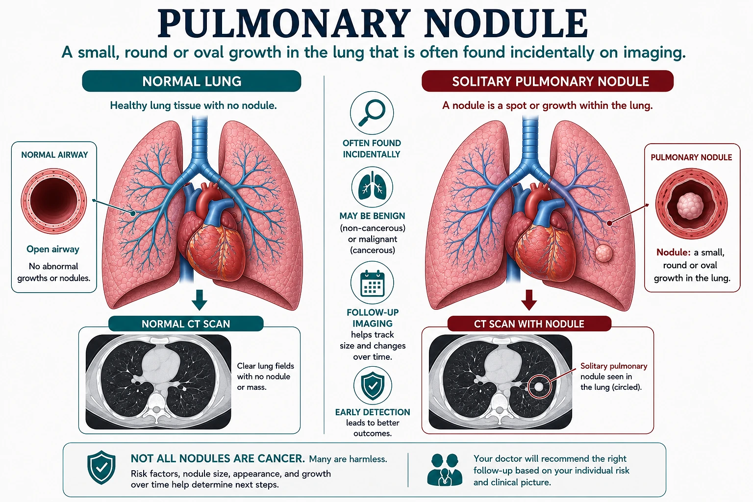
A small, rounded spot in the lung, usually found incidentally on imaging done for another reason. Most pulmonary nodules are not cancer, but evaluation and follow-up are individualized.
In short
A pulmonary nodule is a small, rounded spot found on a chest X-ray or CT scan, usually by accident while imaging is done for something else. Most nodules are not cancer. What happens next (nothing further, periodic follow-up CT scans, additional imaging, or a tissue sample) depends on the nodule's size and appearance together with the patient's age, smoking history, and overall risk, so it's genuinely individualized rather than a single universal rule.
Pulmonary Nodules at a Glance
What It Is
A small, rounded area of tissue in the lung, typically under 3 centimeters, usually discovered incidentally on imaging.
How Common
Nodules are a frequent incidental finding; the majority turn out to be benign, especially smaller ones.
How It's Found
Most often on a chest X-ray or CT scan ordered for an unrelated symptom, or on a dedicated lung cancer screening CT.
What Determines Next Steps
Nodule size, edges and internal composition, solid versus subsolid appearance, and the patient's age and smoking history together.
Key Takeaways
- A pulmonary nodule is a small, rounded spot in the lung tissue, most often found incidentally on a chest X-ray or CT scan done for an unrelated reason.
- The large majority of pulmonary nodules are not cancer; many represent old, healed infection or other benign processes.
- Nodule size, edges and shape, and whether a nodule is solid or subsolid all factor into how it's assessed, but no single feature tells the whole story on its own.
- Recommendations are individualized: the same size nodule can reasonably lead to different follow-up plans for different patients based on age, smoking history, and overall risk.
- Comparing a new nodule to any prior imaging is one of the most valuable steps in assessing whether it's stable or new, when older scans are available.
- Periodic follow-up CT scans (surveillance) are a common, evidence-based approach for many nodules, though not every nodule is managed the same way.
Symptoms
Typical Presentation
- No symptoms at all in most cases, which is why nodules are usually found incidentally rather than because of how a patient feels
- Discovered on a chest X-ray or CT scan ordered for an unrelated reason, such as a cough, chest pain, or a pre-surgical workup
- Discovered on a dedicated low-dose CT scan performed for lung cancer screening in an eligible current or former smoker
- Occasionally accompanied by symptoms if the nodule is large or located near an airway, though this is the exception rather than the rule
Common Questions After a New Nodule Finding
I was just told I have a lung nodule. Does that mean I have cancer?
No, not usually. Most pulmonary nodules are not cancer; many represent something entirely benign, such as scar tissue from an old, healed infection or a small cluster of inflammatory tissue that has been sitting quietly, unchanged, for years. That said, a real evaluation is still worthwhile in every case, because a minority of nodules do turn out to be something that benefits from being caught and addressed early.
Why didn't I notice any symptoms?
This is expected. Most pulmonary nodules cause no symptoms at all, which is exactly why they're usually found incidentally, on imaging done for something else entirely, rather than because of how a patient feels.
Do I need a biopsy right away?
Usually not. Biopsy isn't the default first step for most nodules, since many are reasonably managed with imaging alone. A recommendation for biopsy generally reflects a specific reason, such as higher-risk features or growth on follow-up imaging.
What should I bring to my appointment?
Any prior imaging that shows the same area of the lung, if it exists, even from years earlier or a different health system. Comparing a new nodule to older scans is one of the most valuable steps in assessing whether it's stable or new.
Will I need more scans after this one?
Often, yes. Periodic follow-up CT scans, known as surveillance, are a common and evidence-based next step for many nodules, though not every nodule is managed the same way, and some need no further imaging at all.
What Can a Pulmonary Nodule Represent?
Old, Healed Infection
Scar tissue from a prior infection, such as histoplasmosis, tuberculosis, or a resolved pneumonia, is one of the most common causes of a benign nodule, particularly in regions where these infections are more common.
Granuloma
A small cluster of inflammatory tissue that forms in response to infection or inflammation and can persist, unchanged, for years without causing any problem.
Benign Tumor
Growths such as a hamartoma, a common non-cancerous lung tumor, can appear as a nodule with a characteristic look on imaging.
Inflammatory or Autoimmune Nodule
Certain inflammatory conditions can produce nodules in the lung as part of a broader disease process elsewhere in the body.
Infection in Progress
An active or recent infection can sometimes appear as a nodule-like finding that resolves or changes on follow-up imaging.
Primary Lung Cancer
A minority of nodules represent an early lung cancer, which is why risk assessment and, when appropriate, follow-up imaging matter even though most nodules are not this.
Metastatic Disease
Less commonly, a nodule can represent spread from a cancer elsewhere in the body, usually considered when a patient has a known cancer history.
Risk Factors
- Nodule sizeLarger nodules carry more statistical concern, though size alone never determines the outcome
- Current or past smoking historyOne of the most significant modifiers of how a given nodule is approached
- AgeRisk of malignancy in a nodule rises with age, particularly beyond the fifth and sixth decades
- Nodule edges and shapeSmooth, well-defined edges are generally more reassuring than irregular, spiculated, or lobulated borders
- Solid versus subsolid appearanceSolid and subsolid (ground-glass or part-solid) nodules are assessed and followed differently
- Growth on prior imagingA nodule that has grown compared to an older scan carries more concern than one that has been stable for years
- Personal or family history of lung cancerAdds to overall risk assessment alongside imaging features
- Occupational or environmental exposuresHistory of exposure to radon, asbestos, or certain other inhaled substances can factor into overall risk
- Nodule location and numberA single nodule is generally evaluated differently than multiple nodules, which can suggest a different underlying process
Why Evaluation Still Matters
Most Nodules Are Not Cancer
This is the most important starting point for any patient told they have a nodule, and it remains true even though a careful evaluation is still worthwhile.
Early Identification Matters When Cancer Is Present
In the minority of nodules that do represent an early lung cancer, identifying and addressing it while it's small and localized is associated with better outcomes.
Unnecessary Anxiety Is a Real Cost
A structured, individualized approach helps avoid both under-evaluation of a concerning nodule and over-testing of a nodule that's very likely benign.
Avoiding Unneeded Procedures
Appropriate risk assessment helps identify which nodules can be safely watched with imaging alone, sparing many patients an invasive test they don't need.
When Should I Talk to a Pulmonary Specialist?
- A nodule newly identified on a chest X-ray or CT scan
- A prior nodule that has grown in size on a follow-up scan
- A nodule with irregular, spiculated, or lobulated edges noted by the radiologist
- A nodule found in a patient with a significant current or past smoking history
- A nodule found alongside unexplained weight loss, coughing up blood, or persistent chest symptoms
- Uncertainty about whether prior imaging is available for comparison
- A recommendation for follow-up imaging that hasn't been scheduled
What Is a Pulmonary Nodule?
The Basic Definition
A pulmonary nodule is a small, rounded area of tissue within the lung, generally defined as smaller than 3 centimeters across. Most pulmonary nodules are not cancer. Many represent something entirely benign, such as scar tissue from an old, healed infection or a small cluster of inflammatory tissue that has been sitting quietly, unchanged, for years.
Why Evaluation Still Matters
Being told “it’s probably nothing” isn’t the same as being told nothing further needs to happen. A real evaluation is still worthwhile in every case, because a minority of nodules do turn out to be something that benefits from being caught and addressed early.
Nodules larger than about 3 centimeters are generally described using different terminology (a “mass”) and are approached with a different, more urgent framework, since size alone shifts the balance of likely causes considerably. This page focuses specifically on the smaller nodule that most patients are actually dealing with.
How Pulmonary Nodules Are Usually Found
Pulmonary nodules are discovered in two main ways, and understanding which pathway applies is useful context on its own.
Incidental Discovery
The first, and by far the most common, is incidental discovery: a chest X-ray or CT scan ordered for an unrelated reason, such as a cough, chest pain, or a pre-surgical workup, happens to show a nodule that wasn’t the reason for the scan. This is genuinely common; imaging technology has become sensitive enough that small nodules turn up far more often than they used to, mostly reflecting better detection rather than a true rise in how often nodules occur.
Screening Discovery
The second pathway is intentional: lung cancer screening. Current guidance recommends annual low-dose CT screening for eligible current and former smokers in a defined age and smoking-history range, since this population carries a meaningfully higher baseline risk. A nodule found through screening is expected and planned for, and the screening program includes a structured follow-up pathway.
Both pathways lead to the same underlying question: given this specific nodule’s characteristics and this specific patient’s risk profile, what happens next?
Size and Morphology: What They Do and Don’t Tell Us
Size
Nodule size is one of the most heavily weighted factors in how a nodule is assessed. As a general pattern, larger nodules carry more statistical concern than smaller ones. But size by itself never tells the whole story, and treating it as the single deciding factor is one of the more common oversimplifications patients run into after being told they have a nodule.
Shape, Edges & Internal Features
Radiologists and physicians also look closely at a nodule’s morphology, its shape and edges. Smooth, well-defined, rounded borders are generally more reassuring than irregular, spiculated (spiky), or lobulated edges, which can suggest a more actively growing process. Internal features matter too, such as calcium in a pattern typical of an old healed process, which can itself be a strongly reassuring sign.
None of these features works in isolation. A radiologist’s description of a nodule’s size and appearance is one input into a broader assessment that also weighs the patient sitting in front of the physician, which is exactly why the same imaging report can lead to different recommendations for different people.
Solid Versus Subsolid Nodules
One of the more important structural distinctions in nodule evaluation is whether a nodule is solid or subsolid. A solid nodule appears as a uniformly dense spot on CT imaging, similar in density to normal soft tissue. A subsolid nodule has a hazier, less dense appearance, and this category is further split into purely ground-glass nodules (entirely hazy, with no solid component) and part-solid nodules (a mix of hazy and denser tissue).
| Dimension | Solid Nodules | Subsolid Nodules |
|---|---|---|
| Appearance on CT | Uniformly dense, similar to normal soft tissue | Hazier and less dense; purely ground-glass or part-solid |
| Subtypes | One category | Purely ground-glass (no solid component) or part-solid (mixed hazy and dense) |
| Typical Pace of Change | Varies with underlying cause | Often slower, changing over years rather than months, particularly part-solid nodules |
| Typical Follow-Up Length | Matched to the nodule's size and risk profile | Sometimes followed for a longer overall period than a comparably sized solid nodule |
This is a general pattern rather than a fixed rule; it’s one more reason a nodule’s specific classification, not just its size, shapes what follow-up looks like.
What a Nodule Can Represent
The range of things a pulmonary nodule can turn out to be is genuinely broad, from old healed infection and benign growths to, less often, an early lung cancer (see the causes listed above). That breadth is exactly why “probably nothing” and “definitely needs treatment” are both oversimplifications on their own, and why evaluation matters even when the overall odds favor a benign explanation.
Risk Assessment: Why It’s Individualized
A physician weighs these factors together, not separately: age, smoking history, family and personal cancer history, relevant occupational or environmental exposures, and the nodule’s own imaging characteristics (the risk factors listed above), asking how likely this particular nodule is to need more than watching for this particular patient.
The practical result: two patients with nodules of nearly identical size and appearance can reasonably receive different recommendations, one needing no further imaging while the other is offered a shorter surveillance interval or additional testing. Neither is wrong; each reflects a different overall risk profile applied to a similar-looking finding. It’s easy to compare notes with a friend or relative who had a “similar” nodule and assume the same plan should apply, when the plan was never based on the nodule alone.
Why Comparing to Prior Imaging Matters So Much
Why Stability Over Time Matters
If a nodule looks essentially unchanged compared to a scan taken years earlier, that stability is one of the single most valuable pieces of information available, in many cases more informative than any additional test that could be ordered now. A nodule that has been present and unchanged for two or more years is generally considered very unlikely to represent an active cancer, since most lung cancers grow measurably over that kind of timeframe.
How To Get Prior Imaging
This is precisely why gathering prior imaging, or having a previous facility send records to a new physician, is worth real effort. Patients sometimes don’t realize they have an old scan of the same area, whether from a hospital visit years earlier, a different health system, or an unrelated emergency room evaluation. Locating that old imaging can occasionally resolve a nodule question outright, or at minimum shorten how long surveillance needs to last.
CT Surveillance
For many nodules, especially those with lower or intermediate risk features, the recommended next step is periodic follow-up imaging rather than an immediate additional test. This approach, often called CT surveillance, means having a repeat CT scan at an interval chosen for that specific nodule and patient, then comparing the new scan to the prior one to check for growth, stability, or resolution.
Surveillance is a genuinely evidence-based, mainstream approach, not a wait-and-see compromise. The large majority of nodules followed this way turn out to be stable and are ultimately dropped from further monitoring once enough time has passed without change.
What Fleischner-Style Frameworks Provide
Well-known frameworks, including guidance published by the Fleischner Society, describe general size- and appearance-based starting points for how surveillance intervals might be structured. These frameworks are genuinely useful and widely referenced in pulmonary practice, but they were explicitly designed to be a starting point for clinical judgment, not a substitute for it.
Why They're Adjusted To The Individual Patient
The guidance builds in adjustments for patient risk: a physician applying it modifies the general framework based on smoking history, age, family history, and the nodule’s specific features, rather than following a single size-to-interval table as though it applies identically to everyone.
A low-risk nodule in a non-smoker may be managed less intensively than the framework’s starting point suggests, while a higher-risk patient with an identical-sized nodule may reasonably be followed more closely. A specific “if this size, then this interval” claim found online is worth treating with some skepticism unless it’s paired with that same individualization.
PET/CT and Biopsy: When More Than Surveillance Is Considered
PET/CT
For nodules where imaging and risk assessment together leave meaningful uncertainty, particularly larger solid nodules, a PET/CT scan can help estimate how metabolically active the nodule is. It isn’t ordered reflexively for every nodule: smaller and many subsolid nodules aren’t typically well suited to it, and a physician selects it specifically when it’s likely to change the plan.
Biopsy (Tissue Sampling)
When risk assessment and imaging features together suggest a meaningful likelihood of malignancy, or a definitive diagnosis is needed to guide treatment, a tissue sample (biopsy) may be recommended. Several approaches exist, chosen based on the nodule’s size, location, and the patient’s overall health; the specific technique is decided individually rather than following a single standard procedure.
Biopsy isn’t the default first step for most nodules: many are reasonably managed with imaging alone, and a recommendation for it generally reflects a specific reason, such as higher-risk features, growth on follow-up, or a patient’s preference for a definitive answer over continued watching.
Bringing It Together
A pulmonary nodule is, most of the time, a reassuring finding once properly evaluated: a small, rounded spot that turns out to be old scar tissue, a healed infection, or a benign growth. That reassurance is real and worth stating plainly.
At the same time, evaluation matters, because the minority of nodules that represent something more significant are far better addressed early than late, and there’s no way to sort a specific nodule into the reassuring or concerning category without looking closely at its own features alongside the patient’s broader risk profile. The steps below summarize how those pieces come together in practice.
- 01Nodule IdentifiedFound incidentally on imaging done for another reason, or on a dedicated lung cancer screening CT.
- 02Size & Appearance ReviewedSize, edges and shape, and solid versus subsolid appearance are assessed on the imaging itself.
- 03Compared to Prior ImagingWhen older scans are available, the nodule is compared to check whether it's stable or new.
- 04Individual Risk WeighedAge, smoking history, family history, and relevant exposures are factored in alongside the imaging findings.
- 05Personalized Next StepNo further imaging, CT surveillance, additional imaging such as PET/CT, or a tissue sample, decided together with a physician.
That combination, not any single number or test result, is what determines the right next step. Two patients can reasonably receive two different plans for what looks, on paper, like a similar nodule, and that’s expected, not a sign that one of them received the wrong advice.
Pulmonary Nodule Evaluation in Frisco, Texas
Patients in the Frisco, Texas area evaluating a newly identified pulmonary nodule, including gathering prior imaging for comparison and building an individualized follow-up plan, can be seen through VitalAir Sleep & Lung Center's pulmonary evaluation services. The clinical approach described on this page applies regardless of where that evaluation takes place; what a local visit adds is simply a physician working through these specific factors for a specific patient's nodule.
Patient Questions
What is a pulmonary nodule?
A pulmonary nodule is a small, rounded spot of tissue within the lung, generally defined as smaller than 3 centimeters across. It's almost always found incidentally, meaning it turns up on imaging performed for an unrelated reason rather than because it caused symptoms on its own.
Is a pulmonary nodule usually cancer?
No. The large majority of pulmonary nodules are not cancer. Many represent old, healed infection, a benign growth, or scar tissue from prior inflammation. That said, a minority of nodules do represent an early lung cancer, which is why a physician's evaluation of the specific nodule's size, appearance, and the patient's individual risk factors is still worthwhile rather than assuming either extreme.
What size nodule is dangerous?
There isn't a single size cutoff that separates a dangerous nodule from a safe one. In general, larger nodules carry more statistical concern than smaller ones, but size is only one factor among several, including the nodule's edges and shape, whether it's solid or subsolid, whether it has grown compared to prior imaging, and the patient's age and smoking history. A small nodule in a higher-risk patient can sometimes warrant more attention than a somewhat larger nodule in a lower-risk patient. This is exactly why nodule follow-up is individualized rather than governed by a single size threshold that applies to everyone.
How are pulmonary nodules usually found?
Most are found incidentally, on a chest X-ray or CT scan ordered for something unrelated to the nodule itself, such as evaluating a cough, chest pain, or preparing for an unrelated surgery. A growing number are also found on dedicated low-dose CT scans performed specifically for lung cancer screening in eligible current or former smokers, where finding a nodule is an expected and planned-for possibility rather than a surprise.
What's the difference between a solid and a subsolid (ground-glass) nodule?
Solid nodules appear as a uniformly dense spot on CT imaging. Subsolid nodules, which include purely ground-glass nodules and part-solid nodules, have a hazier, less dense appearance, sometimes with a solid component mixed in. These two categories behave differently and are generally followed on different schedules, since subsolid nodules, particularly part-solid ones, can carry meaningful risk even when they grow very slowly over long periods of time.
Why does comparing a new nodule to old scans matter so much?
If a nodule looks essentially identical in size and appearance on a scan taken years earlier, that stability over time is one of the most reassuring pieces of information available, often more useful than any single test. Conversely, a nodule that has grown compared to a prior scan is treated with more concern regardless of its current size. Bringing prior imaging (or asking a prior facility to send records) is genuinely one of the most useful things a patient can do before a nodule evaluation.
What is CT surveillance for a pulmonary nodule?
CT surveillance means having periodic follow-up CT scans, spaced at intervals chosen based on the nodule's size, appearance, and the patient's overall risk, to see whether the nodule stays the same, grows, or resolves. It's a common and evidence-based approach for many nodules, particularly those with lower to intermediate risk features, because most benign nodules simply don't change over time.
How often will I need a follow-up scan?
There is no single interval that applies to every patient. Professional frameworks such as the Fleischner Society guidelines describe general starting points based on nodule size and whether it's solid or subsolid, but those frameworks are explicitly meant to be adapted based on a patient's individual risk factors, the specific appearance of the nodule, and a physician's clinical judgment. Two patients with a similarly sized nodule can reasonably be given different follow-up plans.
When is a PET/CT scan used for a pulmonary nodule?
A PET/CT scan is sometimes used for larger solid nodules that carry an intermediate likelihood of malignancy after standard imaging, to help estimate how metabolically active the nodule is. It's a tool used selectively rather than for every nodule; smaller nodules and many subsolid nodules aren't typically well suited to this particular test.
When is a biopsy needed for a pulmonary nodule?
A tissue sample is generally considered when a nodule's imaging features and a patient's risk profile together suggest a meaningful chance of malignancy, when a nodule has grown on follow-up imaging, or when a diagnosis is needed to guide treatment decisions. It isn't the default first step for most nodules, since many can be reasonably followed with imaging alone.
Can smoking history change how a nodule is managed?
Yes, significantly. A current or past smoking history is one of the strongest modifiers of how a nodule is approached, since it raises the baseline likelihood that a given nodule could be malignant. Two patients with an identical-looking nodule can be given meaningfully different recommendations based on this factor alone.
Should I see a pulmonologist for a pulmonary nodule?
A pulmonology evaluation is a reasonable step for most newly identified pulmonary nodules, particularly to interpret the specific imaging findings, gather any prior scans for comparison, assess individual risk factors, and determine an appropriate, individualized follow-up plan rather than guessing based on size alone.
Sources
Guidelines and Professional Societies
- Fleischner Society guidelines for management of incidentally detected pulmonary nodules on CT images, describing size- and appearance-based follow-up frameworks intended to be adapted to individual patient risk.
- American College of Chest Physicians (CHEST) evidence-based clinical practice guideline on evaluation of individuals with pulmonary nodules, including risk stratification for likelihood of malignancy.
- American Thoracic Society — patient and clinician educational resources on pulmonary nodules and lung cancer screening.View source
Government and Regulatory Sources
- United States Preventive Services Task Force recommendation on lung cancer screening with low-dose CT in eligible current and former smokers.
- National Institutes of Health / National Cancer Institute — overview of pulmonary nodule evaluation and lung cancer risk factors.View source