Pulmonary Medicine
Medically reviewed by Varun Halani, MD · August 14, 2026
Pleural Effusion
Fluid accumulation in the pleural space, covering the transudative vs. exudative distinction, common causes from heart failure to infection to malignancy, evaluation with imaging and thoracentesis, and management approaches.
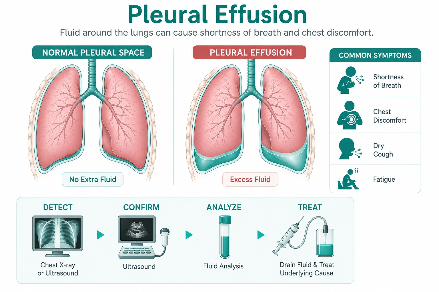
In short
A pleural effusion is a buildup of fluid in the pleural space, the thin space between the lung and the chest wall. Effusions are classified as transudative or exudative based on testing the fluid itself, most often using Light's criteria, and that classification narrows down the likely cause: transudative effusions typically come from a systemic pressure or protein imbalance such as heart failure, cirrhosis, or nephrotic syndrome, while exudative effusions point to a more local process such as infection, malignancy, or pulmonary embolism. Evaluation combines imaging with thoracentesis, a needle procedure that samples the fluid, when the cause is not already clear. Management ranges from treating the underlying condition to therapeutic drainage or, for complicated or recurrent effusions, a longer-term drainage approach.
Pleural Effusion at a Glance
What It Is
Fluid buildup in the pleural space, the thin space between the lung and chest wall. A finding with many possible causes, not a single disease.
How It's Classified
Transudative or exudative, based on testing the pleural fluid itself, most often using Light's criteria.
How It's Evaluated
Imaging confirms the effusion; thoracentesis, needle sampling of the fluid, usually determines the specific cause.
How It's Treated
Ranges from treating the underlying condition to therapeutic drainage, with a chest tube or longer-term catheter for complicated or recurrent effusions.
Key Takeaways
- A pleural effusion is fluid accumulation in the pleural space, the thin space between the lung and the chest wall; it is a finding with many possible causes, not a diagnosis in itself.
- Pleural fluid is classified as transudative or exudative, most often using Light's criteria, and this distinction meaningfully narrows the list of likely causes.
- Heart failure, cirrhosis, and nephrotic syndrome are the classic causes of transudative effusions; infection (parapneumonic effusion), malignancy, and pulmonary embolism are among the most common causes of exudative effusions.
- Imaging confirms an effusion, and thoracentesis, sampling the fluid with a needle, is typically needed to determine the cause when it is not already apparent from the clinical picture.
- A parapneumonic effusion that becomes infected (empyema) or is large and complicated typically needs drainage with a chest tube, not antibiotics alone.
- Management always addresses the underlying cause; large or recurrent effusions may also need therapeutic thoracentesis or a longer-term drainage approach such as an indwelling pleural catheter.
Symptoms
Common Symptoms
- Shortness of breath, especially with a larger effusion
- A dull, aching, or sharp chest pain that may worsen with a deep breath (pleuritic pain)
- A dry cough
- A feeling of chest heaviness or fullness
Symptoms That May Point to the Underlying Cause
- Fever and a productive cough, suggesting infection
- Leg swelling and difficulty breathing while lying flat, suggesting heart failure
- Unintentional weight loss, suggesting malignancy
- Abdominal swelling and a history of liver disease, suggesting cirrhosis
Could This Be a Pleural Effusion?
My chest imaging showed fluid around my lung. What does that mean?
It means you have a pleural effusion, fluid in the space between the lung and the chest wall. On its own, this is a finding rather than a diagnosis. The next step is usually determining whether the fluid is transudative or exudative, which helps identify the underlying cause.
I have heart failure and I'm more short of breath than usual. Could this be a pleural effusion?
Yes, this is a common pattern. Heart failure is one of the most common causes of a transudative pleural effusion, and worsening shortness of breath in someone with known heart failure is a reasonable reason to check for one.
I had pneumonia and I'm still short of breath and running a fever. Could I have developed a complication?
Possibly. A parapneumonic effusion, fluid that develops alongside pneumonia, is a recognized complication, and if it becomes infected (empyema) it typically needs drainage rather than antibiotics alone. Persistent fever or breathlessness after starting pneumonia treatment is worth reevaluating promptly.
I have a history of cancer and new shortness of breath. Should I be concerned about a pleural effusion?
It's reasonable to raise this with your physician. Malignant pleural effusion is one of the more common causes of an exudative effusion in someone with a known or new cancer diagnosis, and new or worsening breathlessness deserves evaluation rather than being assumed to be unrelated.
What Causes a Pleural Effusion?
Transudative Causes
Result from a systemic pressure or protein imbalance rather than direct pleural disease. Heart failure, cirrhosis, and nephrotic syndrome account for the large majority of transudative effusions.
Exudative Causes
Result from a more local process affecting the pleura or nearby lung tissue. Infection (parapneumonic effusion), malignancy, and pulmonary embolism are among the most common.
Other Recognized Causes
Autoimmune disease, recent chest surgery or trauma, certain medications, and, less commonly, other systemic conditions can also produce a pleural effusion.
Risk Factors
- Heart failureOne of the most common causes of a transudative pleural effusion.
- CirrhosisCan cause a transudative effusion, often called hepatic hydrothorax, typically on the right side.
- Nephrotic syndromeA less common but recognized cause of transudative effusion related to low blood protein levels.
- Pneumonia or another lung infectionA leading cause of exudative effusion (parapneumonic effusion).
- Active or prior cancerMalignant pleural effusion is one of the more common causes of exudative effusion, particularly with lung, breast, and lymphatic cancers.
- Pulmonary embolismA recognized, sometimes overlooked cause of exudative effusion.
- Recent chest surgery, trauma, or a chest procedureCan cause a reactive or blood-tinged effusion.
- Autoimmune diseaseConditions such as rheumatoid arthritis and lupus are recognized, less common causes.
Why an Untreated or Undiagnosed Effusion Matters
Breathing Mechanics
A large effusion compresses the adjacent lung, reducing its ability to expand fully and increasing the effort required to breathe.
Delayed Diagnosis of the Underlying Cause
Because a pleural effusion is a sign of an underlying process rather than a stand-alone disease, not identifying that cause promptly can delay treatment of conditions such as infection, malignancy, or heart failure.
Risk of Infection Progressing to Empyema
A parapneumonic effusion that is not adequately drained can progress to a frankly infected collection (empyema), which is harder to treat and carries greater risk.
Recurrence
Effusions caused by an ongoing process, such as active malignancy or poorly controlled heart failure, can reaccumulate after drainage unless the underlying cause is also addressed.
When Should I Talk to a Pulmonary Specialist?
- New or worsening shortness of breath, particularly if it is progressive
- Pleuritic chest pain, a sharp pain that worsens with a deep breath or cough
- Fever, productive cough, or other signs of infection alongside breathlessness
- A known diagnosis of heart failure, cirrhosis, or cancer with new respiratory symptoms
- An incidental finding of pleural fluid on imaging obtained for another reason
- Persistent symptoms or fever despite treatment for pneumonia
What a Pleural Effusion Is
Where Fluid Accumulates
The pleural space is a thin, normally fluid-filled space between the lung and the chest wall, containing only a small amount of lubricating fluid under normal conditions. A pleural effusion is an abnormal accumulation of additional fluid in that space, which can compress the adjacent lung and reduce how fully it can expand.
Why It's a Sign, Not a Diagnosis
A pleural effusion is not itself a disease; it is a physical finding that can result from a wide range of underlying conditions, from heart failure to infection to cancer. Because the appropriate treatment depends entirely on what is causing the fluid, identifying the underlying cause, rather than simply confirming that fluid is present, is the central task of evaluation.
Transudative vs. Exudative: The Central Distinction
Once a pleural effusion is confirmed, the fluid itself is generally tested to classify it as transudative or exudative, most often using a set of laboratory comparisons called Light’s criteria. This single distinction meaningfully narrows the list of likely causes and shapes what further evaluation is needed.
Transudative vs. Exudative Effusions
| Dimension | Transudative Effusion | Exudative Effusion |
|---|---|---|
| Underlying Mechanism | A systemic pressure or protein imbalance; the pleura itself is not directly diseased | A local process that increases the leakiness of the pleura or nearby lung tissue |
| Light's Criteria Result | Does not meet any of the three exudative thresholds | Meets at least one of three thresholds: pleural fluid-to-serum protein ratio above 0.5, pleural fluid-to-serum LDH ratio above 0.6, or pleural fluid LDH above two-thirds the upper limit of normal serum LDH |
| Common Causes | Heart failure, cirrhosis (hepatic hydrothorax), nephrotic syndrome | Parapneumonic effusion and empyema, malignancy, pulmonary embolism, autoimmune disease |
| Typical Next Step | Often managed by treating the underlying condition, with monitoring rather than extensive further pleural testing | Usually requires further evaluation of the fluid itself, such as cytology or culture, to pin down the specific cause |
Light’s criteria are highly sensitive for identifying exudative effusions, though they can occasionally misclassify a transudative effusion as exudative, for example in someone with heart failure who has already been treated with diuretics before the fluid is sampled. Results are always interpreted together with the overall clinical picture rather than in isolation.
Common Causes in Context
Heart failure, cirrhosis, and nephrotic syndrome account for the large majority of transudative effusions, reflecting the shared mechanism of a systemic pressure or protein imbalance rather than disease of the pleura itself. When heart failure is suspected as the cause, an echocardiogram is usually the first test used to evaluate heart function. In more complex or ambiguous cases, particularly when distinguishing a cardiac cause from another explanation would meaningfully change the treatment plan, right heart catheterization can directly measure the pressures responsible for fluid buildup.
On the exudative side, infection, malignancy, and pulmonary embolism are the three causes most often actively searched for once an effusion is classified as exudative, since each requires a distinctly different treatment path.
How a Pleural Effusion Is Evaluated
The Diagnostic Pathway
- 01History and ExamSymptoms, known medical conditions such as heart failure or cancer, and recent illnesses such as pneumonia are reviewed for clues to the likely cause.
- 02Imaging ConfirmationA chest X-ray usually confirms the effusion first; ultrasound helps confirm free-flowing fluid and safely guides a thoracentesis, and a CT scan is added when more anatomic detail is needed.
- 03ThoracentesisFluid is sampled with a needle and sent for cell count, protein and LDH levels (Light’s criteria), Gram stain and culture, and cytology, along with other targeted tests based on the suspected cause.
- 04Fluid ClassificationLight's criteria classify the fluid as transudative or exudative, which narrows the differential and guides what additional testing, if any, is needed.
- 05Additional Testing as NeededDepending on the suspected cause, this can include bronchoscopy when an airway lesion or lung cancer is suspected, an echocardiogram for suspected heart failure, or imaging for pulmonary embolism.
- 06Treating the Underlying CauseOnce identified, treatment is directed at the underlying condition, alongside drainage of the fluid itself when clinically appropriate.
Chest CT imaging is particularly useful when an effusion is loculated, or walled off into pockets, or when an underlying lung abnormality such as a mass needs to be characterized more fully than a chest X-ray allows. Bronchoscopy is not a routine part of every pleural effusion evaluation, but it becomes relevant when an airway abnormality or lung cancer is suspected as the underlying cause.
Parapneumonic Effusion and Empyema
A parapneumonic effusion, fluid that develops alongside pneumonia, follows a spectrum from simple to complicated. A simple parapneumonic effusion is typically sterile and often resolves as the underlying pneumonia is treated with antibiotics. A complicated parapneumonic effusion, or a frankly infected collection known as empyema, generally does not resolve with antibiotics alone and requires drainage, most often with a chest tube, since infected or thickened fluid does not reliably clear on its own. Persistent fever, worsening breathlessness, or a lack of improvement despite appropriate antibiotic treatment for pneumonia are reasons to reevaluate for this possibility.
Malignant Pleural Effusion
Malignant pleural effusion occurs when cancer cells directly involve the pleura, either from a primary lung cancer or from another cancer that has spread there. It is diagnosed by sending fluid obtained through thoracentesis for cytology, and sometimes requires a pleural biopsy when cytology alone is not conclusive. Because malignant effusions often reaccumulate as long as the underlying cancer is active, management frequently moves beyond a single thoracentesis toward an indwelling pleural catheter or pleurodesis, chosen based on the specific clinical situation, expected prognosis, and patient preference.
Treatment and Management Approaches
Treatment always starts with, and often centers on, the underlying cause: optimizing heart failure therapy, treating an infection, or managing cirrhosis, as appropriate. For effusions that are large, symptomatic, infected, or of unclear cause, a procedure directed at the fluid itself is also part of care.
Diagnostic and therapeutic thoracentesis is often the first procedural step, both to obtain fluid for testing and, when a large effusion is causing shortness of breath, to relieve that symptom directly. When fluid is infected, thickened, or loculated, as can occur with empyema, chest tube drainage provides more complete and sustained removal than thoracentesis alone. For effusions that are expected to recur, most often malignant effusions, an indwelling pleural catheter or pleurodesis reduces how often repeat drainage procedures are needed going forward.
Getting an Accurate Diagnosis
Because a pleural effusion can be caused by conditions as different as heart failure, infection, and cancer, and because the right treatment depends entirely on which of these is responsible, a thorough evaluation that includes fluid analysis is generally the right approach rather than assuming a cause based on the clinical picture alone.
For patients in the North Dallas-Fort Worth area, the pulmonary team at VitalAir Sleep & Lung Center in Frisco, Texas offers evaluation for unexplained shortness of breath or an abnormal chest imaging finding, which can be an appropriate starting point when a pleural effusion is discovered or suspected.
Patient Questions
What is a pleural effusion?
A pleural effusion is an abnormal buildup of fluid in the pleural space, the thin space between the lung and the chest wall that normally contains only a small amount of lubricating fluid. It is a finding that can result from many different underlying conditions, not a diagnosis in itself.
What is the difference between a transudative and an exudative pleural effusion?
This is the central classification used to evaluate a pleural effusion. Transudative effusions result from a systemic pressure or protein imbalance, most often heart failure, cirrhosis, or nephrotic syndrome, and the fluid itself looks relatively dilute on testing. Exudative effusions result from a more local process, such as infection, malignancy, or pulmonary embolism, that increases the leakiness of the pleura itself. Distinguishing the two, most often with Light's criteria, meaningfully narrows the likely cause and shapes what further testing is needed.
What are Light's criteria?
Light's criteria are a set of laboratory comparisons between the pleural fluid and the blood, used to classify an effusion as exudative. An effusion is considered exudative if any one of three thresholds is met: a pleural fluid-to-serum protein ratio greater than 0.5, a pleural fluid-to-serum LDH ratio greater than 0.6, or a pleural fluid LDH more than two-thirds the upper limit of normal for serum LDH. Light's criteria are highly sensitive for exudative effusions, though occasionally a transudative cause, such as heart failure treated with diuretics, can be misclassified as exudative, which is one reason results are interpreted alongside the full clinical picture.
What is a parapneumonic effusion?
A parapneumonic effusion is a pleural effusion that develops alongside pneumonia or another lung infection. Many parapneumonic effusions are simple and resolve with antibiotic treatment of the underlying infection, but a complicated parapneumonic effusion, or a frankly infected one (empyema), typically requires drainage with a chest tube rather than antibiotics alone.
What is a malignant pleural effusion?
A malignant pleural effusion occurs when cancer cells directly involve the pleura, either from a lung cancer or from another cancer that has spread to the pleura. It is one of the more common causes of an exudative effusion in someone with a known or newly suspected cancer, and diagnosis typically involves sending fluid obtained by thoracentesis for cytology, examination of the fluid for cancer cells.
What is a thoracentesis, and is it painful?
Thoracentesis is a procedure in which a physician inserts a needle through the chest wall into the pleural space to remove fluid, guided by ultrasound to improve safety and accuracy. It is performed under local anesthesia, and most patients describe pressure or brief discomfort rather than significant pain. The fluid collected is sent for testing to help determine the cause of the effusion, and removing a large volume can also relieve shortness of breath.
Do all pleural effusions need to be drained?
No. A small effusion, particularly one clearly explained by a treatable cause such as heart failure, is sometimes managed by treating the underlying condition and monitoring the effusion rather than draining it immediately. Larger, symptomatic, infected, or diagnostically unclear effusions are more likely to need thoracentesis or another drainage approach.
Can a pleural effusion come back after it's drained?
Yes, particularly when the underlying cause is still active, such as ongoing malignancy or poorly controlled heart failure. For effusions that reaccumulate repeatedly, an indwelling pleural catheter or pleurodesis can reduce how often repeat drainage procedures are needed.
How is right heart catheterization related to a pleural effusion?
When heart failure is suspected as the cause of a transudative effusion, an echocardiogram is usually the first test used to evaluate heart function. In more complex or ambiguous situations, particularly when distinguishing a cardiac cause from another explanation meaningfully changes the treatment plan, right heart catheterization can directly measure the pressures responsible for fluid buildup.
Sources
Guidelines and Professional Societies
- British Thoracic Society. BTS Guideline for Pleural Disease. Thorax, 2023.View source
- Feller-Kopman DJ, et al. Management of Malignant Pleural Effusions. An Official ATS/STS/STR Clinical Practice Guideline. American Journal of Respiratory and Critical Care Medicine, 2018.View source
- Colice GL, et al. Medical and Surgical Treatment of Parapneumonic Effusions: An Evidence-Based Guideline. Chest, 2000.
Government and Regulatory Sources
- National Heart, Lung, and Blood Institute. Pleural Disorders: pleurisy, pleural effusion, and pneumothorax.View source
Key Evidence
- Light RW, Macgregor MI, Luchsinger PC, Ball WC Jr. Pleural Effusions: The Diagnostic Separation of Transudates and Exudates. Annals of Internal Medicine, 1972.